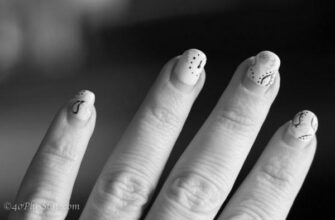
How Can I Get the Kunulae Back to My Fingernails After Anemia?

Your fingernails are made of germinal matrix cells visible through the nail plate. The part of the germinal matrix that is visible is called the lunula. The rest of the matrix is hidden beneath the proximal fold of the nail, known as the eponychium. The germinal matrix cells produce the shine on the nail plate, but only a tiny part of the entire cellular mass is visible.
Anemia

Getting the lunula back to your fingernails after anemia is an important question to ask yourself. Your fingers are essential because they contain signs of various health issues, including iron deficiency. A missing lunula in your hand can signify a thyroid, pituitary gland, or iron deficiency. The largest on your hand, the thumb lunula, is a symptom of an iron deficiency.
There are various causes of lunula loss, including poor nutrition, improper hydration, and exposure to certain chemicals. While it’s hard to trace the exact cause, the condition can be an early warning sign of dangerous situations. Symptoms of such a condition include difficulty breathing, chest pain, and unusual swelling. If your lunulae are prominent or completely absent, seek medical help immediately.
The condition can be caused by anemia, an iron deficiency, or an underlying disease. Anemia is usually caused by not absorbing enough iron from our foods. However, other health problems, including kidney failure, can also cause anemia. The most common of these is iron deficiency. Treatment for anemia is easy and can be achieved by eating iron-rich foods. Iron-rich foods include lean meats, spinach, raisins, and beans.
If your fingernails become discolored, they may signify a rare cirrhosis disease. A person with this condition often experiences yellow nails due to poor blood circulation and may suffer from thyroid disease and sunburn. The lunulae will be discolored in some patients, but this is not a cause for concern.
Diabetes

If you wonder, “How to get the lunulae to return to my fingers in diabetes?” you are in good company. This condition can occur because of various health problems, including heart problems, circulatory problems, and heavy metal poisoning. However, there are certain things you should know first before attempting to return your lunulae to your fingers.
Large lunulae may be a symptom of undiagnosed diabetes, a disease that affects the body’s ability to control blood sugar levels. Fluoride, meanwhile, can cause lunulae to turn brown or black. Moreover, silver poisoning is another common cause of discolored or missing lunulae, and it typically produces thick, slow-growing nails. Eventually, the middle portion of the nail may completely disappear, and the entire nail will take on a yellowish appearance.
If you notice a large lunula on your fingernails in diabetes, you should visit your doctor for a proper diagnosis. If the nail bed is pinkish, you might have cirrhosis or a form of kidney failure. Alternatively, you might have a red lunula, which signals heart failure or kidney failure. Sometimes, your nails will turn half-and-half, another symptom of chronic renal failure.
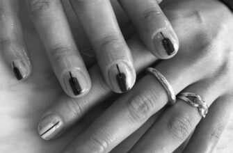
The half-moon-shaped structure at the base of your fingernails is called the lunula. If your fingernails do not have the lunulae, you may have a vitamin deficiency or a medical condition. Your fingernails grow from a pocket beneath the skin called the matrix, which helps new cells push out of the skin.
Silver poisoning

The lunulae, the white region between the nail fold and nail plate, can be a warning sign of a severe medical problem. Besides the nail plate, the lunula on the finger also tells your body about your blood circulation, nutrition status, and organ function. They act as an early warning system for your body, and if they are missing or fading, you should seek medical treatment immediately.
If your lunulae are more significant than your fingernails, you may have a cardiovascular condition. If they are smaller, you may be deficient in some vitamin or mineral. If your lunulae are discolored and appear more significant, you may be suffering from a medical condition, such as cirrhosis, which results in liver scarring. Cirrhosis is the most common cause of lunulae returning to the fingers after silver poisoning. Excess alcohol consumption is also a risk factor.
Some people develop a deep blue lunula. These patches are an early symptom of a severe medical problem called argyria. When exposed to too much silver or other toxic chemicals, the liver can produce too much copper and cause this condition. These bluish patches can also occur on the face and eyes. A lunula may be an early sign of other health conditions, such as anemia or chronic kidney failure.
If you’ve already had silver poisoning and your lunulae are a blue-grey color, you may be suffering from an illness called Wilson’s disease. The disease causes excessive accumulation of copper in the vital organs, which results in symptoms like uncontrolled movements and speech problems. Those who suffer from this condition should seek medical care as soon as possible.
Depression

You’re not alone if you’ve ever noticed a bright white half circle on your thumb. The lunula is part of your nail matrix, but it may be less visible if you’ve damaged the nail plate by over filing it or pushing the cuticle too far back. It may be a sign of anemia, malnutrition, or even a depressive disorder. A person with a high IQ (compared to “normal” people) will have a bright white half circle on their thumb’s base.
There are several ways to restore nail color and function. First, you should examine your fingernails. If they’re discolored, it may signify Wilson’s disease, a rare genetic disorder. In the condition, too much copper builds up in vital organs, including the liver and brain. Other symptoms may include uncontrollable movements, a shortened lifespan, or a fluid buildup in the legs.
In addition to promoting healthy nail growth, lunulae can also tell you if there is a problem with your blood circulation or your nutritional intake. The nails provide an early warning system of a possible medical condition. The absence of lunulae may indicate a minor deficiency, or it could signify a severe health issue.
Anolunula

What is the best way to get the lunulae to grow back on my fingernails? A healthy diet and vitamin E supplements can help. There is no single remedy that will work for everyone, so it is best to consult your doctor before attempting to do it independently. However, if you are desperate and need to see a doctor right away, here are a few things you can do to help the lunulae grow back.
Identify the cause of your condition. Many people have the same problem, but their lack of growth is an underlying health condition or a mistaken manicure. The good news is that there are several ways to get the lunulae back on your fingernails. Read on to learn more. Here are three of them. These tips will help you get your fingernails back to normal!
A blue fingernail is caused by a disease known as Wilson’s disease. This genetic disorder causes excess copper to accumulate in vital organs, including the liver and brain. Some other symptoms of Wilson’s disease include unexplained muscle twitching, uncontrollable movements, problems speaking, abnormal swelling of the legs, and a high fever. If you suspect that you might have a genetic disorder, seek medical advice as soon as possible.
They can also advise you on treatment options.
What Are Nail Structures and Its Parts?

Did you know there are four different layers in the nail? The dorsal layer is two or three cell layers thick and contains the oldest, most brutal cells. The middle layer is one to two cell layers thick and includes the softest, youngest cells. These four layers work together to form the nail. Learn about these layers and what they do for your nails by reading this article. It will help you to understand your nails better and care for them.
Nucleus

The nail matrix comprises the proximal part (called the nail matrix) and the distal part (called the lunula). The nail matrix is commonly considered the source of the bulk of the nail plate, but it also receives contributions from other parts of the nail unit. The contrast between the matrix and the different parts of the nail unit allows for the proper characterization of the nail matrix.
The cellular matrix is the underside of the nail plate, and the cells grow outward toward the fingertip or toe. New cells are pushed out and meet resistance when they emerge from the nail bed. They eventually fuse with the nail plate, where they grow longer and flatter. The keratin protein within these new cells does not contain a nucleus. It is composed of a layer of connective tissue that enables them to grow together.
The proximal 50% of the nail matrix provides 81% of the nail plate’s cells. In comparison, surgery performed to the distal 50% of the matrix is less likely to leave a scar. High-resolution magnetic resonance imaging has helped identify the matrix and the dermal zones below. Baran and Haneke described the layer under the distal matrix as “loose connective tissue with a dense microvascular network.”
The proximal fold is adherent to the nail for a short distance. The cuticle, made of stratum corneum, seals the ungual cul-de-sac and forms a continuous fold with the lateral nail folds. The nail bed extends from the lunula to the hyponychium, presenting parallel longitudinal rete ridges.
Dermis

Understanding the nail’s structure can help you care for your nails and diagnose minor problems. A pin consists of several distinct parts, including the root and the dermis. The heart is an area under the skin called the germinal matrix. It contains blood vessels that supply nutrients to the nail. The dermis, the outer layer, is divided into two main layers, the dorsal and the ventral.
It also contains desmosomes, specialized cell structures that help with cell-to-cell adhesion.
The proximal portion of the nail, called the proximal fold, comprises a thin skin layer surrounding the nail’s base.
The plate, or free edge of the nail, is made of translucent keratin. The vessel has grooves that anchor the fingernail to the nail bed. The nail’s base is connected to the fingertip via an eponychium, or cuticle, between the nail plate and the finger’s skin. It fuses the two structures and provides a waterproof barrier. This area is responsible for hangnails, ingrown nails, and paronychium infections.
Keratinocytes

Autophagy is an essential process in degrading large protein complexes, including keratinocytes. The accessibility of individual proteins outside the cytoskeleton is a primary determinant for autophagy. Deleting the Atg7 gene prevents autophagy, resulting in aberrant accumulation of non-structural proteins in the nails of KO mice. While autophagy is essential for the development of claws, a deficiency in this pathway can result in abnormalities of the keratinocytes.
In the early stages of keratinocyte development, they are offspring of stem cells. These young keratinocytes contain little keratin protein and are cuboidal in shape. As they mature, stem cells push older keratinocytes toward the skin’s surface. As they migrate through the skin, they build up a significant amount of keratin. At this point, the keratinocytes from the first suprabasal cell layer. Further differentiation occurs to form granular keratinocytes.
The nails of WT and KO mice were analyzed using an immunofluorescence method. The WT and KO mice had similar nail structures and iBAQ values. The KO mice had a lower concentration of structural proteins and a higher proportion of non-structural proteins. In contrast, the KO mice had normal-looking nails. These results suggest that the Atg7 gene may affect the differentiation of keratinocytes.
The skin is a barrier to the underlying tissues. The skin is resistant to infection since dead keratinocytes block pathogens from entering. The skin also absorbs minor mechanical damage. Nevertheless, epidermal cells reproduce rapidly and repair skin damage as quickly as possible. The melanocytes produce a pigment called melanin, which absorbs UV light. These cells also protect the underlying tissues from damaging UV light.
Nail matrix

The nails on our hands and feet are made of a matrix, which contains layers of cells that make up the matrix. The matrix is found on the posterior half of the nail bed, beneath the proximal fold. These cells are mitotically active and are considered the fastest dividing skin cells. Despite their importance, the nail matrix is exceptionally delicate. Damage to the matrix may result in broken blood vessels, scarring, or a split nail. Therefore, it is essential to take proper precautions when pulling or pushing back cuticles.
The nail matrix is protected by a fold of skin known as the proximal nail fold. This fold covers the base of the visible nail and is commonly mistaken for the cuticle. The distal portion of the matrix is more vulnerable to disease and damage. It is critical to visit a physician if you suspect these conditions. Luckily, many treatments are available to help you protect your nail matrix.
The nail matrix makes the middle third of the nail plate. If you’ve noticed any signs of leukonychia, you likely have this condition.
The visible portion of the nail matrix is the toenail. The toenail is a crescent-shaped whitish substance. Its shape is determined by the nail matrix and is best seen in the little finger and thumb. The toenail’s color comes from the light reflection in the matrix and nail bed. When the toenail is white, the skin under it is thicker and blocks the pink color of the blood vessels below.
Cuticle

If these layers are damaged, the nails can become ingrown, or hangnails may result. Here are some examples of nail problems.
The nail is a flexible plate made of horny tissue called keratin. It is composed of epidermal cells that originate at the nail root. It is the giant sheet in the human body and contains moderate concentrations of mineral salts such as calcium, sulfur, and potassium. The nail plate is located at the nail base and extends over the hyponychium to form the free edge. The cuticle is the hard, dry skin protecting the nail plate and the nail matrix.
The nail plate consists of translucent keratin. It is made up of several layers of dead cells. This layer provides a rigid but flexible base for the nail plate. The nail plate is joined to the nail bed by connecting ridges. The nail plate separates from the nail bed at the fingertip. The cuticle is the rim of the skin that frames the nail plate and protects the hardening nail. The cuticle helps prevent infections from damaging the nail plate and the underlying tissues.
Basic knowledge of nail anatomy is critical for proper nail care. Knowing this information will help you diagnose minor nail problems and communicate with clients more accurately and professionally. It is also necessary for nail professionals to understand the differences between these three parts. There is a difference between a cuticle and an eponychium. As a nail technician, it is essential to be aware of both legs.